Spectral imaging for cancer identification
(RamanProstate project)
Keywords: prostate, cancer, Raman, imaging, chemometrics
Description:
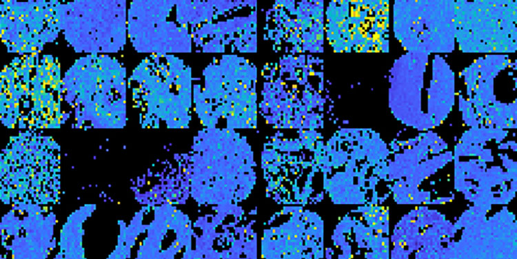
Currently pathologists depend upon staining processes to illuminate important features of tissue samples to increase the clarity of features (e.g. cell nuclei and cytoplasm). Unfortunately the staining process is inconsistent and can be unreliable. Furthermore creating the stains is expensive, laborious and slow. Spectral imaging enables non-destructive analysis of the molecular composition of tissue based on its interaction with light and its potential for identification of various cancer types has been demonstrated.
Raman Prostate investigates the potential of Raman Spectral Imaging, combined with existing methodologies (i.e. staining, digital imaging) using multivariate data analysis to objectively assess prostate tissue. Through collaborations with leading Cancer researchers at UCD’s School of Medicine, data scientists in Dublin Institute of Technology and expert pathologists in Trinity College Dublin and Royal College of Surgeons , this project will develop an integrated data driven approach to enhance traditional prostate tissue evaluation in highlighting and identifying important warning signs of cancer development.
This project is funded the Health Research Board under the HRB Health Research Awards (HRA).