Background: The Human Eye
(By Rahel Krug, student from Berlin, who spent 2 weeks doing an internship at Dr. Breandán Kennedy’s lab (supervised by Dr Nils Ohnesorge))
THE HUMAN EYE
The human eye is a sense organ which is responsible for our vision. It is able to detect light and convert it into electro-chemical impulses. These impulses are then passed on to the brain, where the information is perceived and processed. The eye is made up of various tissue and cells that make differentiation of colours and the depth perception possible. I will go into further detail in the following paragraphs.
EYE STRUCTURE (Figure 1)
The outer layer, which covers most part of the eyeball, is called the sclera. While maintaining the shape of the eye and protecting it, this relatively tough layer also has extraocular muscles attached to it, which are responsible for our eye movement. In front of the iris and the pupil the sclera forms a small, curved, transparent part called the cornea. This is the area where light rays can enter the eye and where they converge. The sclera is covered by a thin, transparent membrane called the conjunctiva, which serves as a protection against damage. The conjunctiva is kept moist and particle free with help from tears coming from our tear glands. The choriod is the second layer of the eye and contains a network of blood vessels to supply nutrients. This layer is deeply pigmented with melanin to prevent light from reflecting inside the eyeball. The iris and the pupil are also part of the choriod. The iris is the muscular, pigmented and thus coloured structure around the pupil. When light enters through the cornea, it passes through the pupil. The amounts of light that enter the interior of the eye are controlled by the iris, making the opening bigger or smaller according to the brightness of the light. The flexible lens is located behind the iris and the pupil. It can change its shape due to ciliary muscles to focus rays of light onto the retina. The above-mentioned retina is the inner layer of the eye and contains a layer of neurons, connected by synapses. The key structures for phototransduction (to convert light into electrical signals) are specialised cells called photoreceptors, which are situated on the outer aspect of the retina. They can be mainly divided into two types: rods and cones (Figure 2).
FIGURE 1 Source: http://www.rexburgvision.com/Eye%20Anatomy.html, 3 May 2014
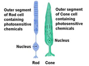 Rods and Cones More rods than cones exist in the human eye. Rods are responsible for sensing contrast, brightness and motion, and respond well to dim light, while cones are capable of performing sharp, central and color vision. The density of cones increases in an area called the macula. Near its center is the fovea, which only consists of cones. There are three different types of cones, who each respond best to different wavelengths of the spectrum of light that reaches the retina. These three types are often referred to as blue (short wavelength), green (medium wavelength) and red (long wavelength) sensitive cones, giving humans the vision which is often referred to as trichromatic. There is also a rarer type of photoreceptors called photosensitive ganglion cells, which don’t contribute to our eyesight directly, but are thought to have a part in circadian rhythms and pupillary reflex. The retinal nerve fibers assemble in the back of the eye, forming the optic nerve, which conducts electrical signals to the brain.
[FIGURE 2. Source: http://clearsci.blogspot.ie/2011/01/sos-save-our-science-seeing-sharks.html, 3 May 2014 ]
At this area, where all the axons converge, there are no photoreceptors. It creates an area that is insensitive to light, the so called blind spot.
The eyeball can also be divided into two main parts which are both filled with fluids. The smaller front section (inside of cornea to front surface of lens) is filled with aqueous humour. This fluid is produced in the ciliary body and exits the eye through the canal of Schlemm. When changes occur in the Schlemm’s canal, which stop the outflow and increase intraocular pressure, an eye disease called glaucoma can result. The larger part in the back of the eye contains a gel-like fluid called vitreous humour, which helps protect the lens. Both fluids are also responsible for maintaining the shape of the chamber of the eyeball.
VISION PERCEPTION
When light enters the eye, it first makes its way through the cornea and then passes through the aqueous humour. The rays are then focused onto the retina by the lens. Here, with the use of rods and cones, phototransduction takes place. Both types of cells contain pigments, made up out of a transmembrane protein called opsin, bound to a group called retinal. Retinal is a form of vitamin A, which explains why vision problems can occur from a lack of this vitamin. Each type of photoreceptor has opsin, although with slightly different amino-acid sequences, which affects the ability to respond to different wavelengths.
PHOTOTRANSDUCTION (ROD VISION)
As shortly mentioned before, phototransduction is the process in which light is absorbed by the pigments in photoreceptor cells and is converted into electrical signals. The pigment molecule that can be found in rods is called rhodopsin and is located in the outer segment of the rod. To understand the process of phototransduction, the situation of the cell when no light reaches the retina, has to be acknowledged. In the dark, the rod cell has a resting potential of about -40 mV. That is more depolarised compared to most neurons in our body. This potential is partly due to open (“leaky”) sodium (Na+) channels, which permit Na+ ions to move into the cell. The channels are kept open by cGMP (Cyclic guanosine monophosphate), which is a molecule that acts as a second messenger. The constant depolarization of the rod cell causes a continual transmitter (glutamate) release from its synaptic terminal, which is important for transmission and communication between rods and interneurons (for example bipolar cells).
When the incoming light is absorbed by retinal, a chain of events occur. The absorption of the photon by the retinal triggers an isomerisation process, which changes the original retinal to all-trans retinal. This also causes the opsin to undergo changes and to activate a G-protein called transducin, which in its inactive state is still bound to GDP. The activation has the effect that transducin then binds to GTP instead and activates an enzyme called phosphodiesterase, which is able to break down cGMP, thus lowering the concentration of these molecules in the outer segment. This drop of cGMP causes the Na+-channels to close, because the molecule cannot bind to the channel receptors anymore. Due to a higher outflow of potassium (Ka+) ions than a Na+ and Ca+ inflow, the cell becomes hyperpolarized (more negative), which ultimately leads to a decreased release of neurotransmitter. (Figure 3) This reduction can either cause a hyperpolarisation or a depolarization in the postsynaptic bipolar cell, thus activating or inhibiting neural pathways. The electric impulses eventually reach ganglion cells and move via the optic nerve to different parts of the brain, where vision can be interpreted. The process of this phototransduction cascade is eventually deactivated by certain proteins that are able to restore the molecules to their inactivated states.
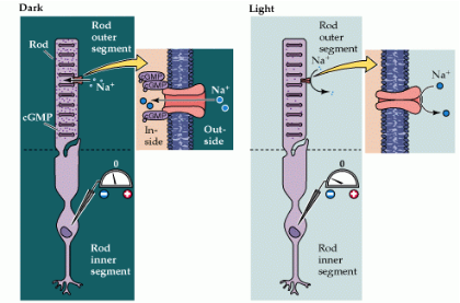
FIGURE 3
Source: http://openwetware.org/wiki/BIO254:Phototransduction, 3 May 2014
CONE VISION
Cones basically go through the same processes as rods, when admitted to light. The difference is that there are three different types of cones, which, as mentioned before, each respond to different wavelengths in different strengths (blue, green and red). (Figure 4) We can therefore see many different shades of colour when these main three are mixed. Cones contain colour pigments instead of rhodopsin, a chemical very similar, except that the opsin part is a little different (photopsin instead of scotopsin). When light of a certain wavelength reaches the cones, all of them will be stimulated, but one will be more stimulated than the others. The cone that responds stronger to a specific wavelength will then have a higher hyperpolarisation, which leads to a stronger decrease in glutamate release. This information of the different photoreceptors can then be compared and processed in different parts of the brain, making our vision of different colours possible. The intensity of colour can be determined in our visual system by comparing how many photoreceptors are responding.
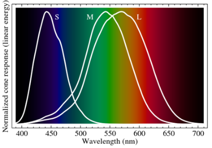
FIGURE 4
Source: http://en.wikipedia.org/wiki/File:Eyesensitivity.svg