This interesting article about a new model of Retinal Vascular Permeability published in Journal of Controlled Release, is the first paper fully funded by this IAPP grant and the second scientific article generated in the project.
A sustained release formulation of novel quininib-hyaluronan microneedles inhibits angiogenesis and retinal vascular permeability in vivo
(opens in a new window)Galvin O, (opens in a new window)Srivastava A, (opens in a new window)Carroll O, (opens in a new window)Kulkarni R, (opens in a new window)Dykes S, (opens in a new window)Vickers S, (opens in a new window)Dickinson K, (opens in a new window)Reynolds AL, (opens in a new window)Kilty C, (opens in a new window)Redmond G, (opens in a new window)Jones R, (opens in a new window)Cheetham S, (opens in a new window)Pandit A, (opens in a new window)Kennedy BN.
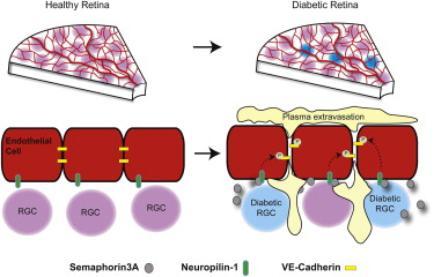
Pathologic neovascularisation and ocular permeability are hallmarks of proliferative diabetic retinopathy and age-related macular degeneration. Current pharmacologic interventions targeting VEGF are effective in only 30-60% of patients and require multiple intraocular injections associated with iatrogenic infection. Thus, our goal is to develop novel small molecule drugs that are VEGF-independent are amenable to sustained ocular-release, and which reduce retinal angiogenesis and retinal vascular permeability. Here, the anti-angiogenic drug quininib was formulated into hyaluronan (HA) microneedles whose safety and efficacy was evaluated in vivo. Quininib-HA microneedles were formulated via desolvation from quininib-HA solution and subsequent cross-linking with 4-arm-PEG-amine prior to freeze-drying. Scanning electron microscopy revealed hollow needle-shaped particle ultrastructure, with a zeta potential of -35.5mV determined by electrophoretic light scattering. The incorporation efficiency and pharmacokinetic profile of quininib released in vitro from the microneedles was quantified by HPLC. Quininib incorporation into these microneedles was 90%. In vitro, 20% quininib was released over 4months; or in the presence of increasing concentrations of hyaluronidase, 60% incorporated quininib was released over 4months. Zebrafish hyaloid vasculature assays demonstrated quininib released from these microneedles significantly (p<0.0001) inhibited ocular developmental angiogenesis compared to control. Sustained amelioration of retinal vascular permeability (RVP) was demonstrated using a bespoke cysteinyl leukotriene induced rodent model. Quininib-HA microparticles significantly inhibited RVP in Brown Norway rats one month after administration compared to neat quininib control (p=0.0071). In summary, quininib-HA microneedles allow for sustained release of quininib; are safe in vivo and quininib released from these microneedles effectively inhibits angiogenesis and RVP in vivo.
(opens in a new window)Link to PubMed