Safer, faster, better ways to image and diagnose disease
Share
Disease diagnosis is often done by taking tissue samples of organs like the liver, kidney or pancreas and running lab tests to spot unwanted changes. Taking these samples – biopsies - can be risky, painful, and time consuming, and it often takes days for a diagnostic result to come back from the lab.
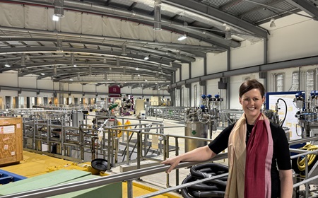
Conway Fellow, Dr Nicola Fletcher is working on new technology that will facilitate quicker, safer, less painful, and more precise micro-biopsies. These micro-biopsies, she said, will involve use of a tiny needle, ten times smaller than current biopsy needles, and yield much faster results.
In parallel, Dr Fletcher is working on game-changing, ‘world’s first’ imaging technology to picture disease-induced changes in individual cells.
Dr Fletcher is a virologist and her love of viruses dates back to her undergraduate days studying science at the University of Limerick.
“I was like a child in a sweet shop in the practical classes, growing viruses in cells and eggs. We grew flu in an egg. I thought it was completely amazing.”
Her skill and enthusiasm for viruses were such that she was offered a job straight after graduating and went to work in the Irish Equine Centre.
“They [viruses] are tiny pieces of DNA wrapped in protein, but can shut down entire countries and bring us to a standstill. I still think it’s incredible.”
It was no surprise when she decided to do a PhD in virology, at UCD, on the subject of the Feline Immunodeficiency Virus. She worked at the Veterinary School in Glasgow after her PhD, and did postdoctoral research at the Medical School in Birmingham before doing a veterinary degree in UCD.
After graduating from UCD in 2018 she worked at the Animal and Plant Health Agency in the UK, studying viruses, TB and prion disease in a range of animals. She applied for an Ad Astra fellowship at UCD but didn’t think she had much change of getting it. She proved herself wrong and began in January 2020.
The plan was to work on the Hepatitis E virus, for which there was no treatment or vaccine, but two months later, the pandemic hit. Her life, and everyone’s, changed, and her focus shifted to SARS-CoV-2 virus, and she was the first to culture the pandemic-causing virus in Ireland.
When the pandemic started to wind down, she pivoted to work on advanced imaging, and in particular using soft x-ray microscopy to study Hepatitis E.
A soft x ray microscope uses low energy x rays to image individual cells. This is done in beautiful detail without having to stain, or prepare cells.
The ability of these microscopes to image cells to see what changes a disease has produced has huge advantages, but they are unable to image tissues.
Dr Fletcher linked up with colleagues at the University of Heidelberg and the University of Warwick to come up with a plan to develop a soft x ray machine that could image tissues. In November 2024, they applied for and secured a European Research Council Synergy Grant for an advanced imaging project - NanoX.
The vision, Dr Fletcher explained, is that, in future, surgeons or pathologists will take a micro-biopsy, freeze the sample, put it into a soft x ray microscope, rotate it, and then image individual cells in detail that wasn’t possible before.
“Any disease will cause structural changes in cells, and we want to image that, and use AI to assign into diseases as there will be massive data sets.”
“At the moment, pathologists take a sample from a patient, fix it, that takes a few days, then slice it up, stain it with dyes and put it on a slide. Then the pathologist uses their years of experience to figure out what is wrong.”
“Traditional pathology is labour-intensive. We want to change that completely.”
The grant started officially on the 1st June 2025 and the focus, initially at least, will be on Hepatitis E, a poorly understood virus for which there is no treatment.
This is all part of an ancient microscopic battlefield, invisible to the eye, where tiny fragments of DNA, coated in protein – called viruses – try to stay ahead of the human immune system, cause disease, even, on occasion, pandemics.
“You have to keep running just to stand still. That’s the constant battle between viruses and the organisms they infect.”
In conversation with journalist, Sean Duke