Image resolution in light microscopy is limited by the wavelength of light and is incapable of resolving structures of less than 200 nm. However, the resolution of electron microscopy limited by biological sample preparation goes beyond 1 nm.
Applications
Research topics include detection of viruses for human patients, inflammation, oncology, cardiovascular biology, morphology of zebrafish retina, bacteriology, food sciences, polymer films for biosensors, nano-particle toxicity in vitro and artificial joints.
Expertise & Services
We can assist with any of the following:
- Experimental strategy, technology choice and planning
- Sample preparation: TEM and SEM
- Image acquisition: TEM and SEM
- Image analysis, including EM tomography
- Training in sample preparation, imaging and image analysis (tailored)
Instrumentation
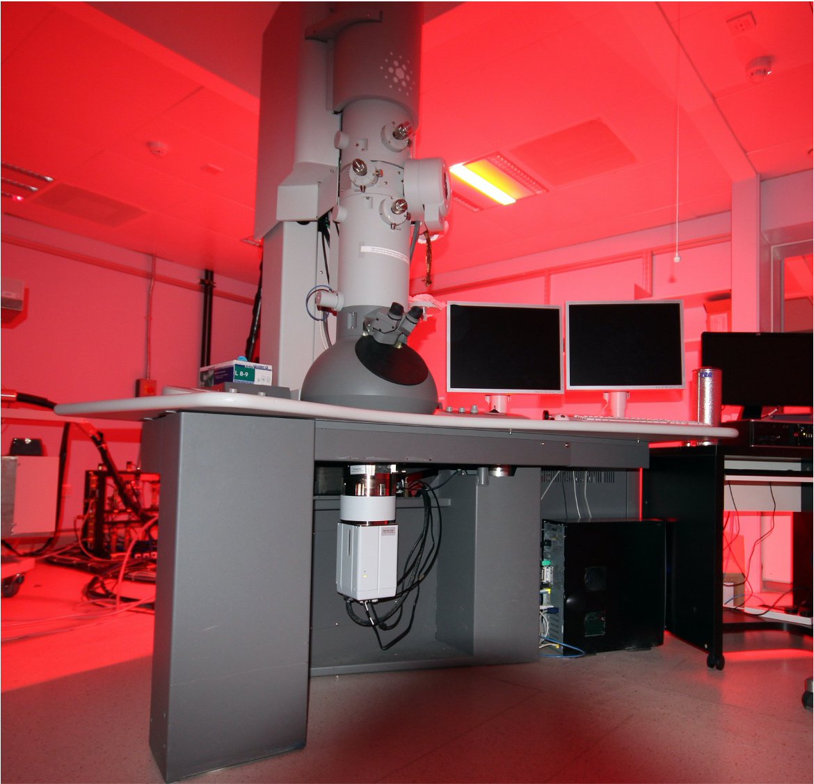
Transmission Electron Microscopy
Transmission electron microscopy (TEM) is used to investigate ultrastructure of thin samples (limited by the penetration of electron beam). Examples include flat cells, nano-particles and biological tissues embedded into polymers. 1 unit available.
Scanning Electron Microscopy
Scanning electron microscopy (SEM) is used to investigate fine structure on surfaces of biological and non-biological objects. 1 unit available.
Support Equipment
Support equipment available for sample preparation includes:
Ultramicrotomes (2) including one suitable for cryo-ultramicrotomy
Fume hood, oven for EPON embedding and other TEM sample preparation instruments
Critical point dryer, gold/carbon coater and other EM sample preparation instruments
Light microscopes for EM sample preparation
.jpg)