 The upper part of the human pelvis, known as the ilium, underwent two major structural innovations during evolution that enabled humans to walk on two legs, a Nature paper reveals. The study lays the developmental and genetic groundwork for the human-defining trait of bipedalism.
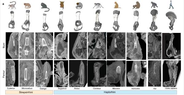
The ilium is the big, flared part of the pelvis that anchors the powerful gluteus maximus muscles that humans use to stay upright. Differences in the illum between humans and other apes are a defining evolutionary feature. However, the developmental processes that led to the unique human ilium shape remain unknown.
The international study led by Dr Terence Capellini, Harvard University, included Niamh Nowlan, Professor of Biomedical Engineering, UCD School of Mechanical and Materials Engineering and Fellow, UCD Conway Institute.
The team used histological, anatomical, and functional genomic approaches to reveal how the structure acquired its unique shape.
One key change involved the direction of cartilage growth. A shift in the orientation of the cartilage growth plate (physis) enabled the ilium to sit perpendicular to the direction seen in ilia from other animals. A second key change involved the process of bone formation. The authors identify timing and spatial differences in the way that bone cells are laid down over the cartilage in the human ilium, compared with non-human primate ilia and human long bones.
Both innovations are interconnected at tissue and molecular levels. Hundreds of regulatory sequences were identified that are active during the development of the human ilium. These show evidence of human evolutionary change, suggesting that key complex, interacting sequences were selected for through time to endow the human pelvis with its unique shape.
 Professor Niamh Nowlan has a research interest in human hip development including from before birth (prenatally). In collaboration with Prof Owen Arthurs, a paediatric radiologist from Great Ormond Street Hospital and University College London Hospitals (UCLH), they assembled a dataset of 3D imaging data from human embryos and foetuses to characterise hip development.
Getting involved in this international research collaboration with Dr Terence Capellini, Chair of the Department of Human Evolutionary Biology and Professor of Human Evolutionary Biology at Harvard University was serendipitous, claims Professor Nowlan.
“When I met Terry, senior author on the paper, he was excited to hear that Owen and I had a dataset that his team could use to characterise how the shape and ossification of the ilium changes over early human development. His team linked this with their extensive data and research on pelvic development in the human embryo and in other animals.”
The research reveals two major developmental genetic innovations shaping the human ilium: a shift in the orientation of the iliac growth plate, which drives its shape and growth and the timing and directionality of iliac bone formation, both of which facilitated further pelvic growth, and the unique formation of the human ilium amongst primates.
“This study reveals the evolutionary shift in morphology of the pelvis that enabled bipedal locomotion, largely through looking at the embryo. Prenatal development of the skeleton is a fascinating subject and there is still so much to learn.
“It reminds me of a famous quote from the renowned 19th century scientist, Viktor Hamburger who said, ‘Our real teacher has been and still is the embryo, who is, incidentally, the only teacher who is always right.’”, said Professor Nowlan.
Read the paper in Nature: https://doi.org/10.1038/
Image captions: Image 1. Iliac ossification between primates including humans; Image 2. Prof Niamh Nowlan.