Research reveals the evolution of human bipedalism in two steps
Monday, 1 September, 2025
Share
How humans first developed the ability to walk on two legs has been revealed after the discovery of critical evolutionary changes which enabled our ancestors to remain standing upright.
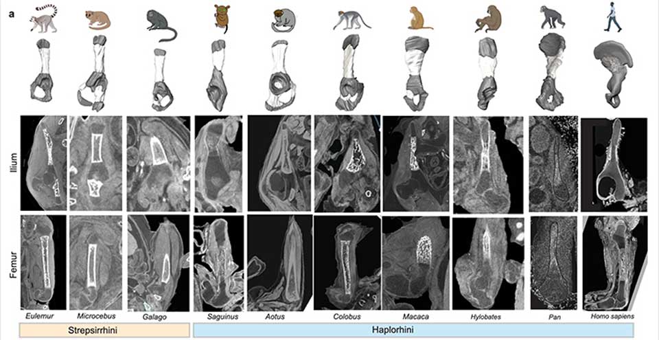
(opens in a new window)A new study published in Nature has identified two major structural changes in the upper part of the human pelvis, known as the ilium, that allowed humans to develop bipedalism and accommodate giving birth to big-brained babies.
The international team behind the research was led by Dr Terence Capellini, Harvard University, and included (opens in a new window)Professor Niamh Nowlan, UCD School of Mechanical and Materials Engineering and Fellow at the UCD Conway Institute.
Their work focused on the ilium – the broad, flared part of the pelvis that anchors the gluteus maximus muscles.
Using anatomical, genomic, and imaging approaches, the team were able to find two key developmental changes that shaped the human ilium into something unique among primates.
First, the orientation of the cartilage growth plate shifted, enabling the bone to sit perpendicular to that seen in other primates.
Second, differences in the timing and direction of bone formation were identified, altering how cells were laid down in the ilium compared to other bones.
Both changes worked together at the tissue and genetic levels, with hundreds of regulatory sequences active during development showing evidence of evolutionary change.
"What we've done here is demonstrate that in human evolution there was a complete mechanistic shift,” said Professor Capellini, Chair of the Department of Human Evolutionary Biology and senior author of the new paper.
“There's no parallel to that in other primates. The evolution of novelty - the transition from fins to limbs or the development of bat wings from fingers - often involves massive shifts in how developmental growth occurs. Here we see humans are doing the same thing, but for their pelves.”
Professor Nowlan, who researches prenatal hip development, contributed to the research a unique dataset of 3D embryo and foetal imaging in collaboration with Professor Owen Arthurs, Great Ormond Street Hospital and University College London Hospitals.
“When I met Terry, senior author on the paper, he was excited to hear that Owen and I had a dataset that his team could use to characterise how the shape and ossification of the ilium changes over early human development,” said Professor Nowlan.
“This study reveals the evolutionary shift in morphology of the pelvis that enabled bipedal locomotion, largely through looking at the embryo… Prenatal development of the skeleton is a fascinating subject, and there is still so much to learn.
“It reminds me of a famous quote from the renowned 19th-century scientist, Viktor Hamburger, who said, ‘Our real teacher has been and still is the embryo, who is, incidentally, the only teacher who is always right’.”
UCD College of Engineering and Architecture
Room 122 & Room 126, UCD Engineering and Materials Science Centre, University College Dublin, Belfield, Dublin 4, Ireland T: +353 1 716 1868 | E: eng.arch@ucd.ie | collegeea@ucd.ie | engarch.research@ucd.ie | Location Map(opens in a new window)All Student Emails should be submitted using our Engineering and Architecture Office Student Connector. This will ensure the fastest and most efficient reply to your email.