Melanocytes: The good, the bad, and the 'surprising'

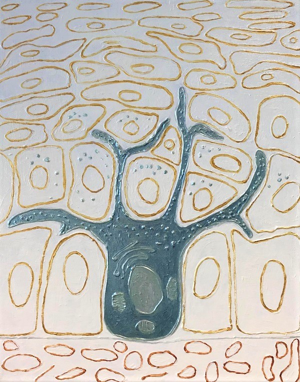
Legend: Artistic representation of a melanocyte cell (in blue) surrounded by keratinocyte cells
(Photo credit: artwork by Christina Kiel)
Melanocytes are melanin pigment-producing skin cells and are responsible for the coloration of skin and hair. Melanocytes reside at the epidermal basement membrane and transport small melanin-containing vesicles (“melanosomes”) through their multiple cellular extensions (“dendrites”) to neighbouring epidermal keratinocytes. Melanin protects the skin from DNA damage by surrounding and guarding the nuclei of keratinocytes. Indeed, melanocytes reach out to – on average – 40 keratinocytes for donation of their melanosomes – qualifying them indeed as extraordinary altruistic cell types in our body.
Malignant melanoma is a highly aggressive cancer, and Ireland has the highest rate of deaths in Europe from this skin cancer (https://www.cancer.ie/cancer-information/melanoma). All melanocytic neoplasms (ranging from benign lesions/naevi to malignant melanomas) originate from melanocytes. For late-stage melanomas and those that are metastasizing, there is no effective treatment and death will occur rapidly, usually within months. It is somewhat surprising that this deadly cancer originates from the ‘friendly’ melanocyte cells, which normally in skin divide only infrequently and that represent a minor cell type in the epidermis (5-10% of the cells in the basal layer). We now know that UV radiation from the sun is a major risk factor – especially with a history of sunburns during childhood. Indeed it is estimated that 60–70% of cutaneous malignant melanomas are caused by UV radiation exposure. It is unfortunate that beach holidays spent carelessly are still very much en vogue.
While melanocytes are generally reduced to either their “good” (melanin producing) or “bad” (melanoma causing) functions, they also have surprising and obscure capabilities. First, they reside in many other human tissues, such as eye, ear, brain, lung, adipose tissue, and even in the heart. Second, melanocytes in non-epidermal skin regions such as hair follicles and hair bulge (the reservoir for melanocyte stem cells) can quite dramatically change their cell fates and identities – over their normal life cycles. For example, during the normal hair growth cycle, melanocytes in hair follicles periodically alter between various cell fates, including proliferating, amelanotic (no production of melanin) and apoptotic fates. These fates are likely controlled by altered extrinsic/cell environmental factors present in the hair follicle niche. A better understanding of how these extrinsic factors, in the context of different cell intrinsic factors (melanoma mutations, melanin content, melanin type, etc), steer cell fates and elucidating the underlying signalling network changes, could potentially provide new targets for drug developments – at least in the long term.
In our research group, we are particularly interested in characterizing and exploiting pathways that are active during normal life-cycles in melanocytes. Specifically, we use co-culture systems to study the impact of microenvironmental cues (e.g. growth factors, hypoxia, inflammation) and cell intrinsic factors (e.g. genetic alterations and mutations, age of melanocytes, melanocytic interaction with keratinocytes) on network alterations and associated cell fates in melanocytes.
Note: May is Skin Cancer/Melanoma Awareness month (https://irishskin.ie/events-calendar/)

About the author:
Christina Kiel is a Science Foundation Ireland-funded Principal Investigator at the UCD School of Medicine in UCD Charles Institute of Dermatology and UCD Systems Biology Ireland. Her group page is available here: http://www.ucd.ie/sbi/people/groups/kielgroup/.

