Easy Single Cell Proteomics
Over the past decade, there have been remarkable advances in the field of single-cell analysis.
A diverse set of innovative tools has been developed, enabling the high-throughput profiling of important biomolecules in individual cells, which can either be extracted from complex multicellular systems or represent distinct cellular states.
Tissues and organs are composed of functionally specialized cells. This specialization of single cells often arises from the protein networks mediating physiological functions. Yet, our ability to comprehensively quantify the proteins including these networks in single cells has remained relatively limited.
Mass spectrometry (MS) is one of the most powerful techniques to quantify thousands of proteins – proteomes - across many thousands of single cells. It has been increasingly recognised that mass spectrometry based on proteomics for single cell analysis provides pictures of cellular behaviours. The improved understanding of biological systems such as tissue development, cancer progression and cell differentiation can be measured simultaneously at single cell level using MS-approach.
In principle, we would like to know for each single cell, the molecular code of the cell (the genome), the functionality of that cell (the proteome and metabolome), and the connection between the two cells (omics).
This required single cell technology extends from genomics to biological function.
Recent technological advances have brought a suite of single cell toolkits that permit robust and high-throughput quantitation of the genome, transcriptome, proteome and metabolome at single cell level. The integration of quantitative analysis inaugurates the multi-omics age of single cell biology and allows researchers to ask questions from perspectives previously unattainable.
For example, in this perspective, the single cell proteome analysis offers great potential to reveal the biology of cancer metastasis and identify signalling pathways for therapeutic interventions. The characteristics of a single cell proteomic assay include multiplexing capacity, throughput, sensitivity and dynamic range. This requires a unified effort of colleagues with diverse expertise, including instrument engineers, separation scientists, mass spectrometry experts, statisticians, and biologists. The increasing need for characterising protein expression at the single cell level requires a robust approach, scalable and multiplexed. Achievement of this goal requires innovations in both handling techniques, i.e., microtechnology such as microfluidics, for improved reliability and scalability, and analytical tools for enhanced sensitivity and multiplexing capability.
However, the increasing complexity of the high-dimensional single cell datasets requires continuous progress in the development of new analytical strategies and computational tools for gleaning useful biological insights from these data.
A major goal of high-dimensional analysis of single cells is not only to understand the relationships among various conceptual aspects of a cell population, but also to generate testable hypotheses regarding how the heterogeneous population dynamically respond and adapt to various cellular and environmental changes.
It is certain that the single cell tools continue to improve in multiplexing capacity, throughput, sensitivity and quantification, an overarching analytical framework that connects biological questions, experimental designs to data analysis and will eventually transform the practice of biomedical research as well as our understanding in single cell biology.
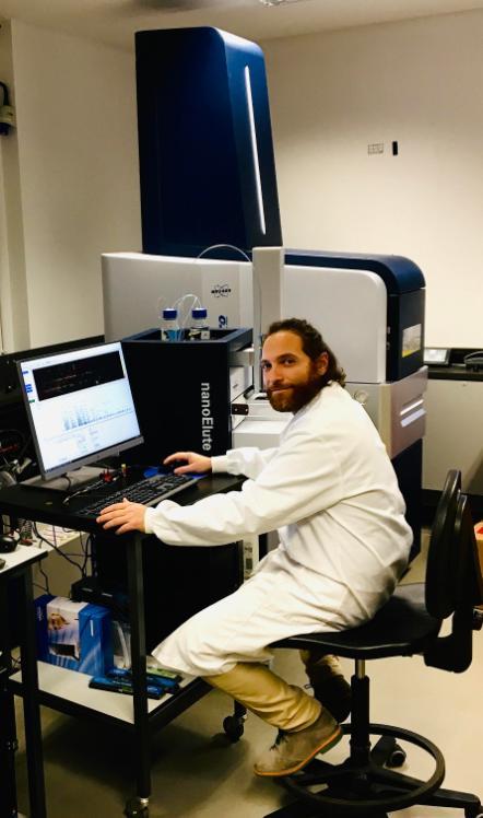
Giorgio at work on one of SBI's Mass Spectrometers.
These notes find inspiration from a recent online conference: “SCP2020 Single-Cell Proteomics Conference”, which brings together many of the pioneers in the fields of proteomics and nano-technologies and aims to create a forum for sharing knowledge, results, and informal discussions in the emerging single cell community.
LINK: https://www.youtube.com/playlist?list=PLHLRxq8iKFsJrLCO47iAe6vh9w0pxLwxV
BRIEF research:
The cell is densely populated by thousands of thousands proteins, which have leading roles in defining the phenotype. Proteins engage in many types of physical interaction to carry out the various functions of a living cell.
Technical developments, in recent years, such as high-resolution high mass accuracy mass spectrometry have significantly increased the power of protein analysis.
However, we are still far from confidently defining how protein dynamics defines cell state. My research exploits methods based on mass spectrometry to identify and quantify thousands of proteins. In particular, I focus on chromatin-related proteins and histone proteins and how their interactors modulate the DNA. Alteration of DNA architecture is mostly driven by two factors:
a) post-translational modifications (PTMs) on histone proteins
b) recruitment of specific chromatin enzymes ensures to change the DNA architecture and ultimate regulate the gene expression.
Today, many clinical chemistry diagnostic laboratories have embraced mass spectrometry as the primary application in multiple researches such as:
- distinguishing cancer and normal specimens, determining cancer aggressiveness and grades, molecular typing of cancer, identifying cancer biomarkers;
- therapeutic drug monitoring from blood, cancer diagnosis, identification of bacteria or virus from throat swab;
- skin and breath metabolite detection;
- Investigation of alteration of gene expression, DNA repair, chromatin compaction, which all together are key factors that define the cell state.

About the Author
Dr. Giorgio Oliviero is an SBI Group Leader whose current research and teaching focuses on proteomics and network analysis using state of the art mass spectrometry instruments. He is originally from Naples, Italy.