What is the “biological identity” of nanomaterials? How do nanoparticles interact with their biological environment? These questions are crucial to the success of nanobiology and nanomedicine.
Nanoparticles pick up proteins on their surface as they enter biological environments such as blood. This protein (biomolecular) corona determines their uptake, transport, final localisation and functional impacts (fate & behaviour), giving them a biological identity. However, how the proteins in the biomolecular corona are presented to cells, in other words, what the cells `see` when they are exposed to nanoparticles remains unclear.
From biochemical methods to imaging techniques, we develop novel approaches to study the surface organization of corona proteins in situ. Of particular interest is the correlation between the physicochemical nature of nanoparticles and the identity, quantity & orientation of the corona proteins. These studies aim to identify protein functional motifs and binding sites that determine specific interactions between nanoparticles and their environment.
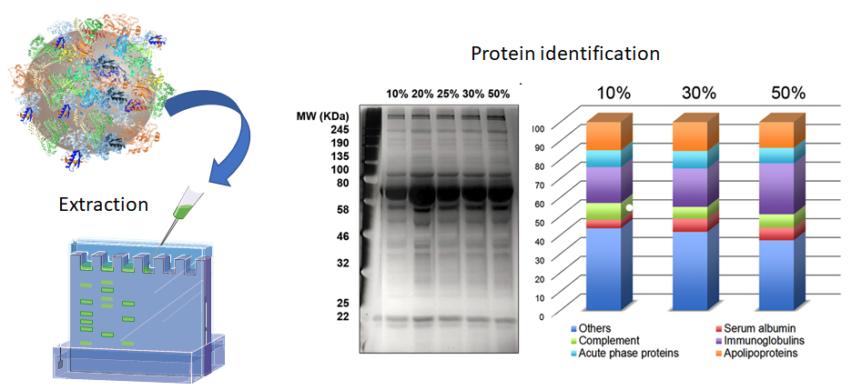
Here SDS-PAGE is used following incubation of nanoparticles with human serum at various concentrations. Distribution of protein groups in human serum corona are analyzed by LC-MS/MS and expressed as protein mass percentage of the total corona proteins. (Lara et al., ACS Nano, 2017)
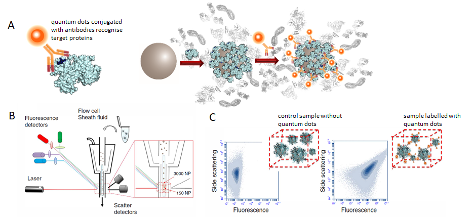
A flow cytometric method has been established to investigate the interactions between nanoparticles and their biological environment. Flow cytometry enables analysis of nanoparticle dispersion and the fluorescence per particle.(A) Quantum dots are functionalized with specific monoclonal antibodies and fluorescently labelled to map out target epitopes of the protein corona. (B) As the number of labelled nanoparticles undergoing laser detection increase, the average scattering properties of the sample change. (C) As a result, the signal-to-noise ratio in the side scattering channel increases enabling the distinction of the signal due to multiple nanoparticles from the instrumental background.(Lo Giudice et al., Nature Communications, 2017)
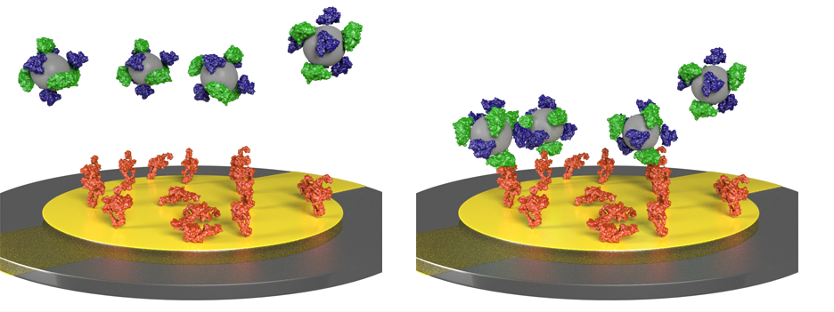
To investigate the recognition of nanoparticles by cell membrane receptors, we are developing a method where we utilise the Quartz Crystal Microbalance instrument. Here, cell membrane receptors are immobilised onto a quartz crystal surface and nanoparticles are presented in a flow. This method allows label-free detection of the interactions between the nanoparticles and the receptors by monitoring the resonance frequency shift of the crystal.(Manuscript in preparation)
Selected publications:
Lara, Sandra, et al. "Identification of receptor binding to the biomolecular corona of nanoparticles." ACS Nano 11.2 (2017)
Herda, Luciana M., et al. "Mapping ofmolecularstructure of the nanoscale surface inbionanoparticles." Journal of the American Chemical Society 139.1 (2016)
Giudice, Maria Cristina Lo, et al. "In situ characterization of nanoparticle biomolecular interactions in complex biological media by flow cytometry." NatureCommunicationss 7 (2016)
Kelly, Philip M., et al. "Mapping protein binding sites on the biomolecular corona of nanoparticles." Nature Nanotechnology10.5 (2015)
Salvati, Anna, et al. "Transferrin-functionalized nanoparticles lose their targeting capabilities when a biomolecule corona adsorbs on the surface." Nature Nanotechnology 8.2 (2013)
Monopoli, Marco P., et al. "Biomolecular coronas provide the biological identity of nanosized materials." Nature Nanotechnology 7.12 (2012)
Centre for BioNano Interactions (CBNI)
School of Chemistry and Chemical Biology, University College Dublin (UCD), Belfield, Dublin 4, D04 N2E5, Ireland. T: +353 1 716 2293 | E: info@cbni.eu | Location Map(opens in a new window)Monday - Friday | 09.00-17.30 Ireland and UK | 10.30-18.30 CET | 00.30-08.30 PST | 17.00-01.30 Japan | 01.00-09.30 PDT