Dean's Lunchtime Clinical Club - 6 February 2018 - João Oliveira
João Oliveira, UCDVH Intern in Equine Clinical Studies, spoke on the 'Conservative Management of a Comminuted Tibial Fracture in a Foal' at the Dean's Lunchtime Clinical Club on 6 February. This case came into the UCDVH last year, and João outlined that tibial fractures are not that common. The 3 month old thoroughbred filly foal had been found acutely non weight bearing lame in the field, and was referred for internal fixation of a tibial fracture to the UCDVH. The UCDVH team carried out a full examination on arrival, and apart from a small focal full skin thickness wound to medial malleolus, everything else was within normal limits. The team examined the radiographs carefully and decided she needed internal fixation; the animal was prepped for surgery.
A CT was then carried out for surgical planning and it was discovered that the animal had a comminuted fracture. Due to the fracture configuration and the high risk of further displacement, it was decided not to proceed with internal fixation of the fracture. Comminution limits the ability of the surgeon to achieve anatomic reconstruction of the tibial cortex, thus further compromising stability. Considerations at that stage were euthanasia or conservative management – the owner decided to go with the latter option. 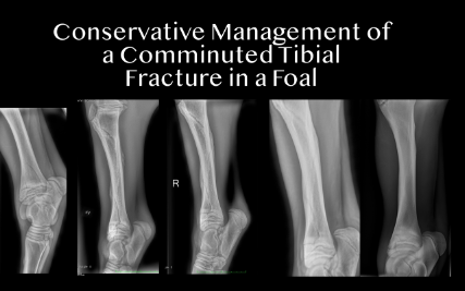
Conservative management of this case involved a tube cast, 2 weeks hospitalisation until the next cast change, box rest, anti-biotics and anti-inflammatories; gastroprotectants were also administered to prevent ulcers. She was monitored intensively and complications during hospitalization were as follows: knuckling into fetlock flexion but will load limb with correct foot placement, self corrected within 3 days of tube casting and able to rise unassisted; contralateral angular limb deformity in the first days; when changing the first cast a small superficial cast sore was noticed on the dorsolateral aspect of the distal third MT bone.
By week 2, the fracture margins were now obvious, there was no displacement of the fracture, and there was secondary bone healing and callous formation. Week 4 brought secondary bone healing with progressive mineralization of the callous and no displacement of the fracture. By week 6, the fracture margins were less obvious, with increased opacity of the callous. Six weeks post fracture the tube cast was replaced with a two layer tube bandage which was removed 48 hours later. The foal then had box rest for 10 days, followed by 20 days cage turnout. At 11 weeks post injury, a gradual increase in turn out duration was introduced. The use of pentosan polysulphate once weekly for 4 weeks, followed by once monthly for a further 4 months, was recommended. 6 months post fracture the foal was walking quite comfortably.
In concluding, João noted that in this case, conservative treatment had a good outcome regarding fracture healing. Intensive care for the first number of weeks is necessary in such cases, as is intensive cast management. The CT was invaluable, and without it, they probably would have gone to surgery and the animal wouldn’t be alive now. This type of case management could be done in a referral Hospital such as the UCDVH, but not in the field, due to the level of care required.